A seizure can be a very scary thing. For the most part, it’s a disorder surrounded by several unknowns. Diagnosing a seizure disorder is usually a process of elimination rather than a definitive diagnosis. Through a battery of tests, one can determine what the seizure is not, easier than telling what exactly it is. The number of disease processes that can lead to seizures is unreal, ranging from hypoglycemia, poisoning, head injury, tumors, stroke, nutrient deficiency, anemia, and the list goes on.1 With so many potential causes of a seizure, I began to wonder if there could also be a structural side of things contributing to these episodes. Could there be something underlying the nutrient deficiency, the blood flow issue, the stroke? Seems like a simple enough question with an underwhelming body of research. In this article I’ve taken a superficial glance at whether or not structural shifting within the spine could be a contributor to these idiopathic seizure disorders.
Let’s Start With Something Simple. What Exactly Is A Seizure?
A seizure is an uncontrolled burst of electrical activity in the brain; the nonstop firing of neurons.1 Basically, the brain misfires. A seizure can last anywhere from seconds to several minutes, there can be a single one of multiple in a row. Often times the animal is left in a daze, confused, lethargic, and sometimes a bit ataxic.
Types Of Seizures:
Therein lies the million-dollar question. With any seizure disorder it is important to know whether the seizure is secondary or primary. For instance, an animal that has a seizure every time it comes in contact with an environmental toxin, is experiencing it on a secondary basis.1 Whereas an animal that has seizures unprovoked and without warning is considered primary. Beyond the initial stages of classification there is another subdivision. Within primary seizures there is grand mal, focal, psychomotor, and idiopathic.
Grand mal seizures sometimes start as focal, and then progress into a much longer and more severe situation. A psychomotor seizure can resemble an odd behavior, such as chasing an object that isn’t really present. These are difficult to identify because many animals do tend to chase imaginary objects or run in circles after their tails. With a seizure, however, the same activity will occur every single time and should last for similar periods. Lastly, idiopathic seizures basically occupy the “we have no idea” category. These are fairly common and do not fit any particular description. Some have hypothesized that these are genetic.2 Seizures can be a result of a big range of diseases, and for the purposes of this article we would like to focus on them being a result of structural abnormality.

It has been suggested that every animal has a seizure threshold, and only some end up crossing over that capacity.3 The same can be said for humans in most walks of life. Every being has a threshold they are born with, and each of us has an innate ability to manage said threshold. When our bodies are compromised, whether it is from physical or mental stressors, those thresholds can be breached and disease expressed. If this is the case for animals, it makes sense that there is very little rhyme or reason as to who will end up being afflicted by seizures, though one might hypothesize an animal in a high stress environment would be more prone to do so.
In my opinion, basic skeletal anatomy may also play a part. If one looks at the shape of a canine skull for instance, it varies between breeds. Those breeds that tend to be afflicted with idiopathic epilepsy (border collies, Australian shepherds, Labrador retrievers, beagles, etc) have very similar skull shapes.1,4 They are all classified as mesocephalic, or moderate skull to snout ratio. Now there are several breeds in the world predisposed to seizure disorders, but perhaps there is something anatomically that is being overlooked. If the skull of an animal related to the cervical spine is within normal limits, what then could be contributing to seizures?
Much like the human spine, animals have a nervous system bathed in cerebrospinal fluid. This fluid acts as a garbage disposal, transporter, shock absorber, and food source for the brain and spinal cord. CSF is produced by the choroid plexus and the ependymal cells.5 It journeys through the brain between ventricles and the subarachnoid space to eventually exit the skull surrounding the spinal cord down to the lumbar cistern.5,6 It allows the cord and brain to move (though minimally) without causing harm to itself. The CSF has a very specific level of protein and nutrients it must maintain in order to keep homeostasis in check. If anything is altered it can be detrimental to the animal.5,6,7
The spine acts as a suit of armor around the spinal cord. The range of motion of the spine allows the spinal cord to move freely without being disturbed. When a structural shift (subluxation) takes place in the spine, limiting the range of motion available, there is disruption to the spinal cord. Specifically, there can be direct pressure placed on the nerves by a disc, by inflammatory fluid, or by the bone itself. Indirect pressure on the nerves and spinal cord may also be present due to the malposition of vertebrae. If a bone is subluxated in one direction, there can be a domino effect. For instance, the dentate ligaments connect the spinal cord to the dura mater, which are then connected by epidural ligaments to the spinal column. When the spinal column is misaligned, the epidural ligaments are stretched and strained, thus transmitting pressures to the dura mater which then alters the normal pressure of the dentate ligament attachments to the spinal cord.
When range of motion is restored through an adjustment, the joint can move freely and the pressure is released off the parties involved. The atlas, or the first cervical vertebrae, plays an important role in the flow of cerebrospinal fluid.6 If the atlas bone were to misalign in a particular direction, the likelihood of the cerebrospinal fluid flow being disrupted in some fashion is highly probable. If it slides up the occipital condyles, the opposite side will have a decrease in the foramen size. Though the decrease may only be millimeters that would be enough to disrupt the flow of fluid.

If the actual change in foramina size didn’t directly disrupt fluid flow, there is also the possibility that the atlas misalignment would cause an increased tension via the dentate ligaments onto the spinal cord and on the blood vessels themselves. This increased tension would raise the pressure necessary to pump blood, etc. The venous system works on hydrostatic pressure, so when there is a rise in said pressure there will be more fluid flowing away from that specific area. With hydrostatic pressure the norm is to flow from areas of high pressure to those of low. Any altering in the pressure will directly alter fluid flow. That disruption as stated previously can have a huge effect on the normal function on the body. In theory, an atlas bone out of place could directly impact the CSF, which could possibly result in a seizure.
The same can be said for the disruption of blood supply to the brain. The brain, though making up a small percentage of total mass, requires an enormous amount of blood.6 There are three main arteries that supply blood to the brain and they run along with the spinal cord. The ventral spinal and the dorsolateral spinal arteries travel through the intervertebral foramina up to the brain where actual nutrient exchange takes place. If somewhere along the way this flow is disrupted by a structural shift in the spine, then the brain could be devoid of necessary nutrients and or oxygen for a period of time, potentially leading up to a seizure. A shift in structure could also affect the sympathetic system as discussed earlier, causing vasoconstriction if some vessels of the brain thus decreasing perfusion to the brain. If blood flow gets backed up and cannot exit the brain in a timely fashion, the arterial blood will have difficulty getting to the brain and once again swelling and potential for seizing exists.
The reason I suggest a possible connection between seizures and a structural shift of that top vertebrae is simple, I’ve had multiple clients have complete resolution of seizures after beginning chiropractic care, and each one of these clients has had a severely subluxated atlas. Similar results have been noted in the literature of humans with seizure disorders. Though the anatomy is definitely different between a biped and a quadruped, the overall concept is quite similar.
References:
http://pets.webmd.com/dog-seizure-disorders
http://www.merckvetmanual.com/pethealth/dog_disorders_and_diseases/brain_spinal_cord_and_nerve_disorders_of_dogs/parts_of_the_nervous_system_in_dogs.html
http://www.canine-epilepsy.com/Why.html
http://biosphera.org/international/images/?pg=dog&i=imagens/anatomia-canina/axis.jpg&d=Dog%20Anatomy:%20Skeleton:%20Axis
https://en.wikivet.net/Cerebral_Spinal_Fluid_-_Anatomy_%26_Physiology
https://en.wikivet.net/Nervous_and_Special_Senses_-_Anatomy_%26_Physiology
http://vetsci.co.uk/2010/02/09/arterial-blood-supply-to-the-brain/
Photo Credit:
yeah yeah yeah…can i go now? via photopin (license)
sweet sweet breath via photopin (license)
Lick via photopin (license)
Awoken In Black And White via photopin (license)
Mily’s tongue via photopin (license)
IMG_0814 via photopin (license)
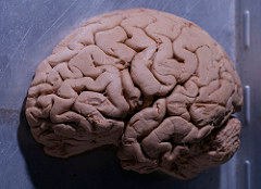
Left Hemisphere, fronal via photopin (license)
science museum via photopin (license)

